Brain Surgery
Evaluation and treatment planning for selected cranial conditions, using advanced microsurgical and endoscopic approaches when clinically appropriate. The recommended surgical approach depends heavily on the specific diagnosis, high-resolution imaging, and individual patient circumstances. Whether addressing complex brain tumors, vascular conditions like aneurysms, or performing delicate cranioplasty reconstructions, we leverage state-of-the-art neurosurgical technology. Our multidisciplinary care ensures that every patient receives a highly tailored treatment plan focused on maximum safety and optimal neurological outcomes.
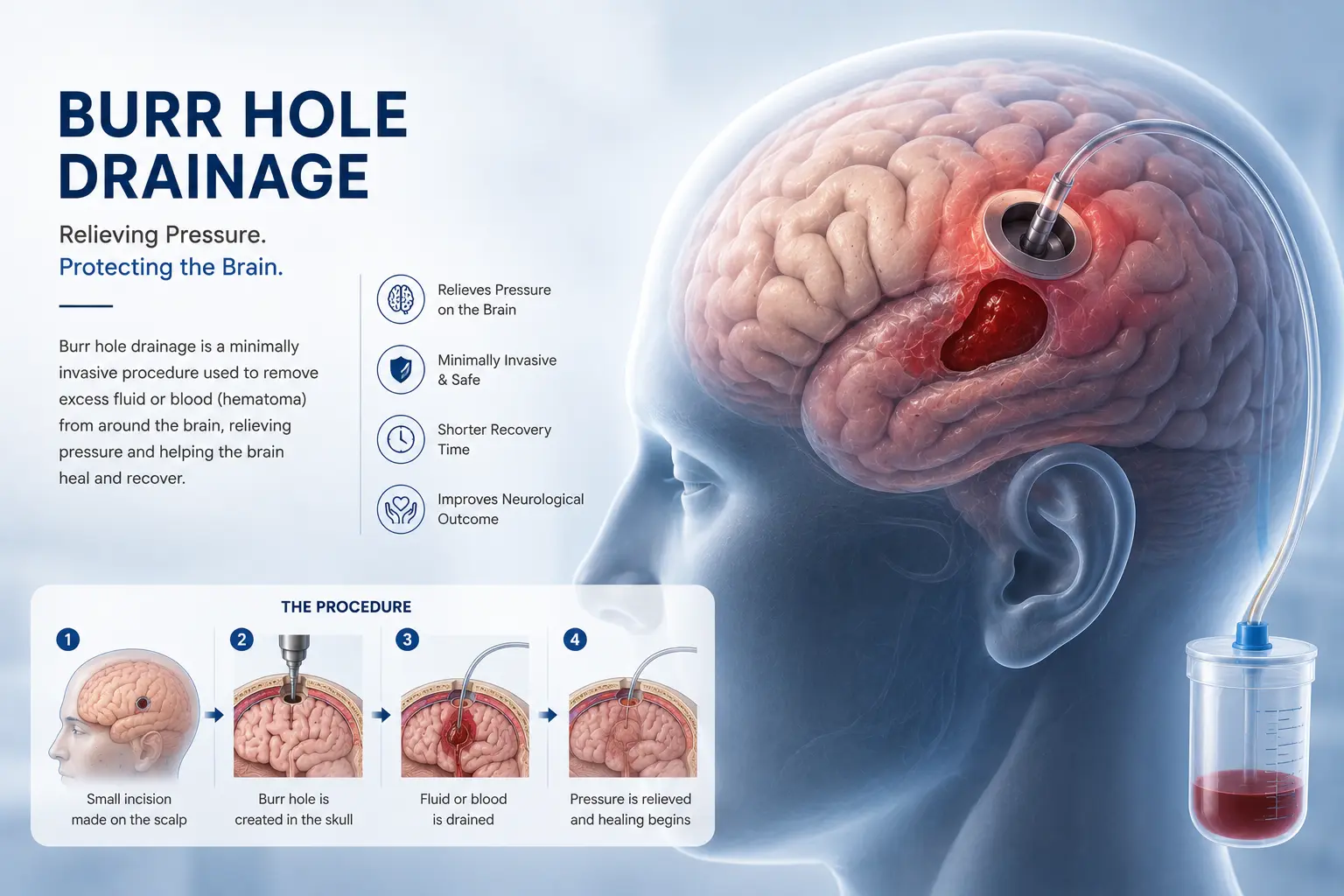
Burr Hole Drainage
A minimally invasive procedure to relieve pressure on the brain by draining fluid or blood. By making a small, precise hole in the skull, surgeons can safely evacuate chronic subdural hematomas or insert monitoring devices, providing immediate relief from life-threatening intracranial pressure while ensuring a rapid recovery.
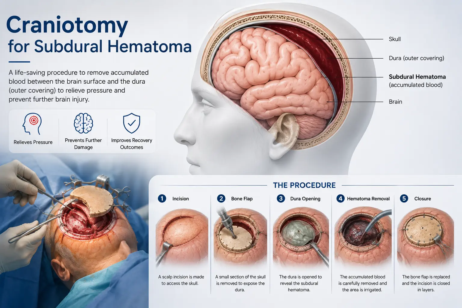
Craniotomy for Subdural Hematoma
Removal of accumulated blood underneath the dura mater to rapidly reduce intracranial pressure. This procedure involves temporarily removing a section of the skull to access and clear acute or complex hematomas that cannot be managed with burr holes alone, meticulously controlling bleeding to prevent permanent brain damage.
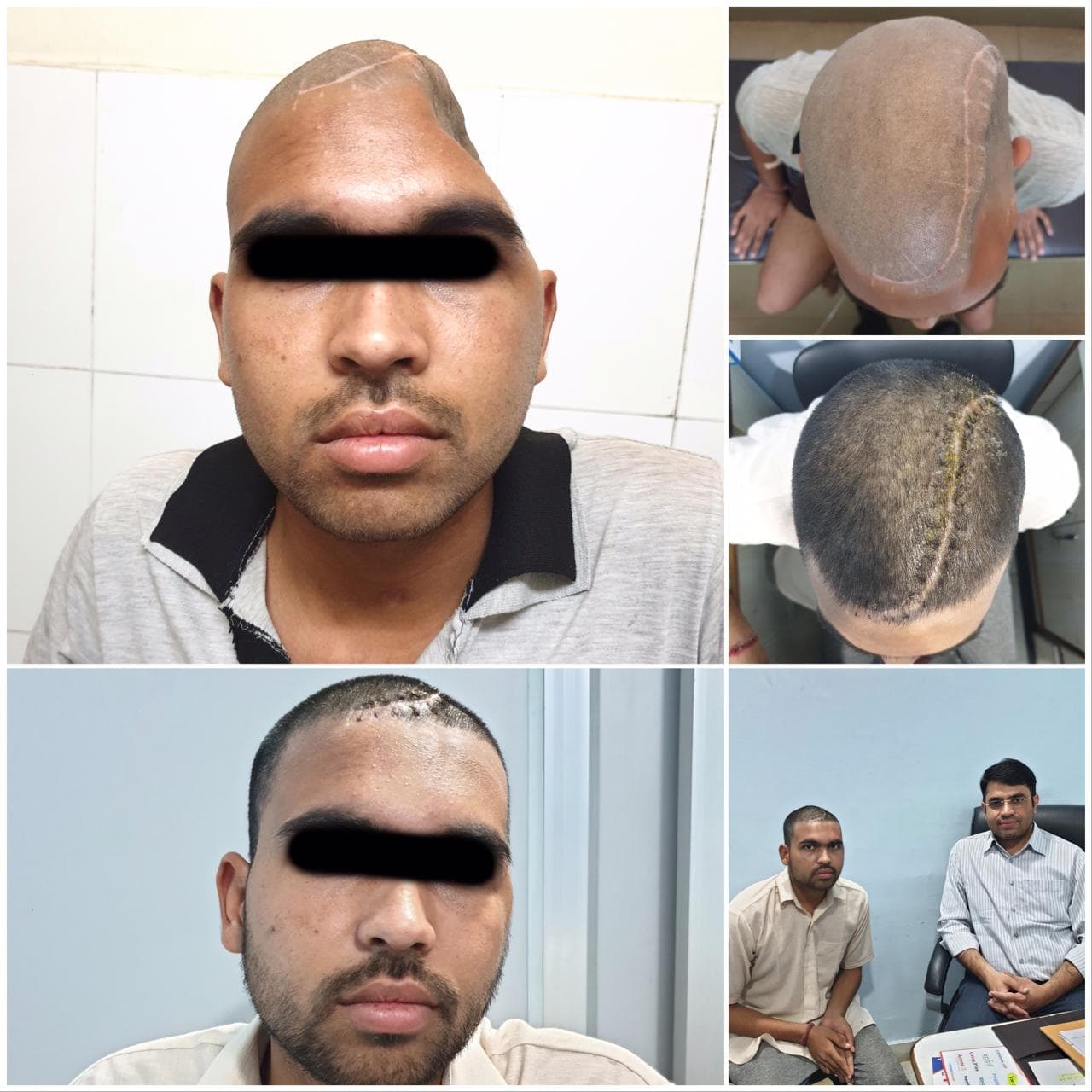
Cranioplasty (3D Printing)/ Patient-Specific Cranial Implant
Reconstruction of skull defects after trauma or previous surgery. When suitable, custom implants may be used as part of a carefully planned reconstruction. Utilizing advanced 3D printing technology, we create patient-specific titanium or synthetic bone flaps that perfectly match the patient's unique cranial contours, restoring both optimal aesthetics and vital brain protection.
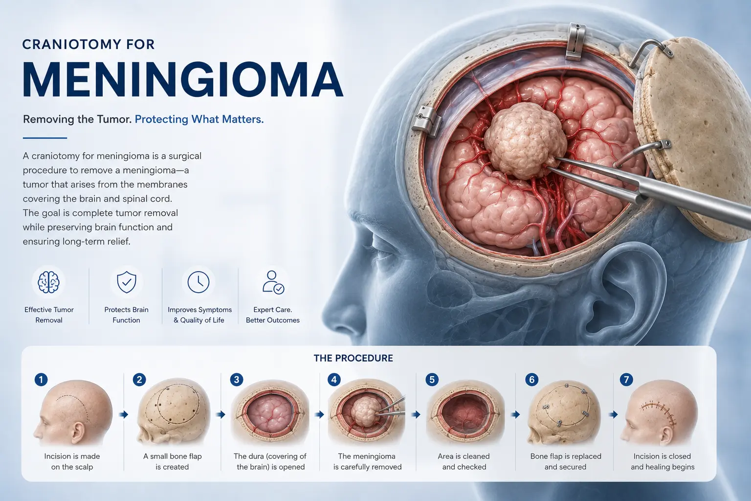
Craniotomy for Meningioma
Surgical removal of a meningioma tumor by temporarily removing a bone flap from the skull. Because these tumors grow from the brain's protective membranes rather than the brain tissue itself, advanced microsurgical techniques allow us to carefully separate the tumor from critical blood vessels and nerves, aiming for complete resection and long-term cure.
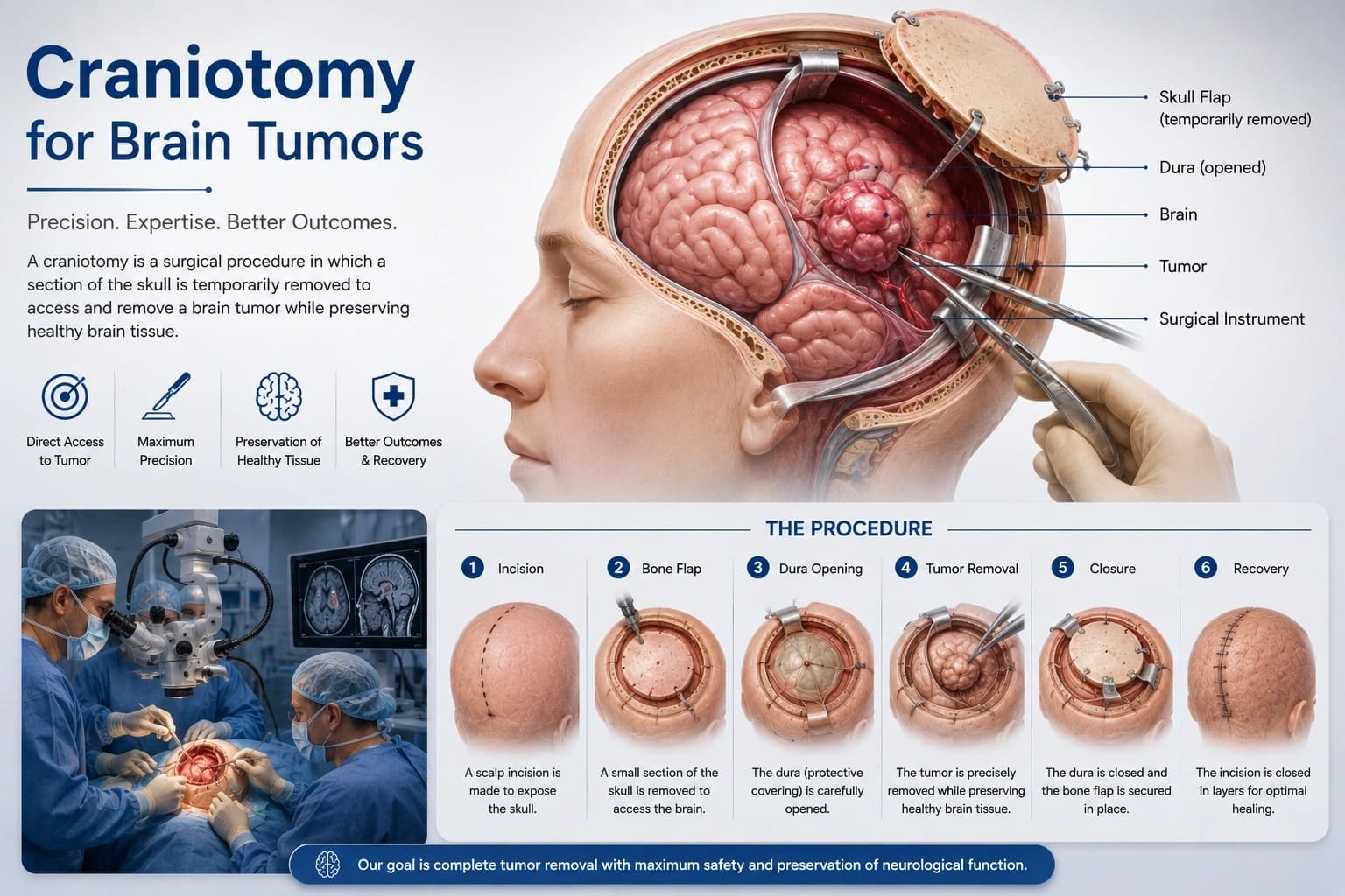
Craniotomy for Brain Tumors
Precise surgical opening of the skull to access and carefully resect brain tumors. Utilizing state-of-the-art neuronavigation, intraoperative imaging, and functional brain mapping, our surgeons safely maximize tumor removal (such as gliomas or metastases) while preserving healthy tissue and essential neurological functions.
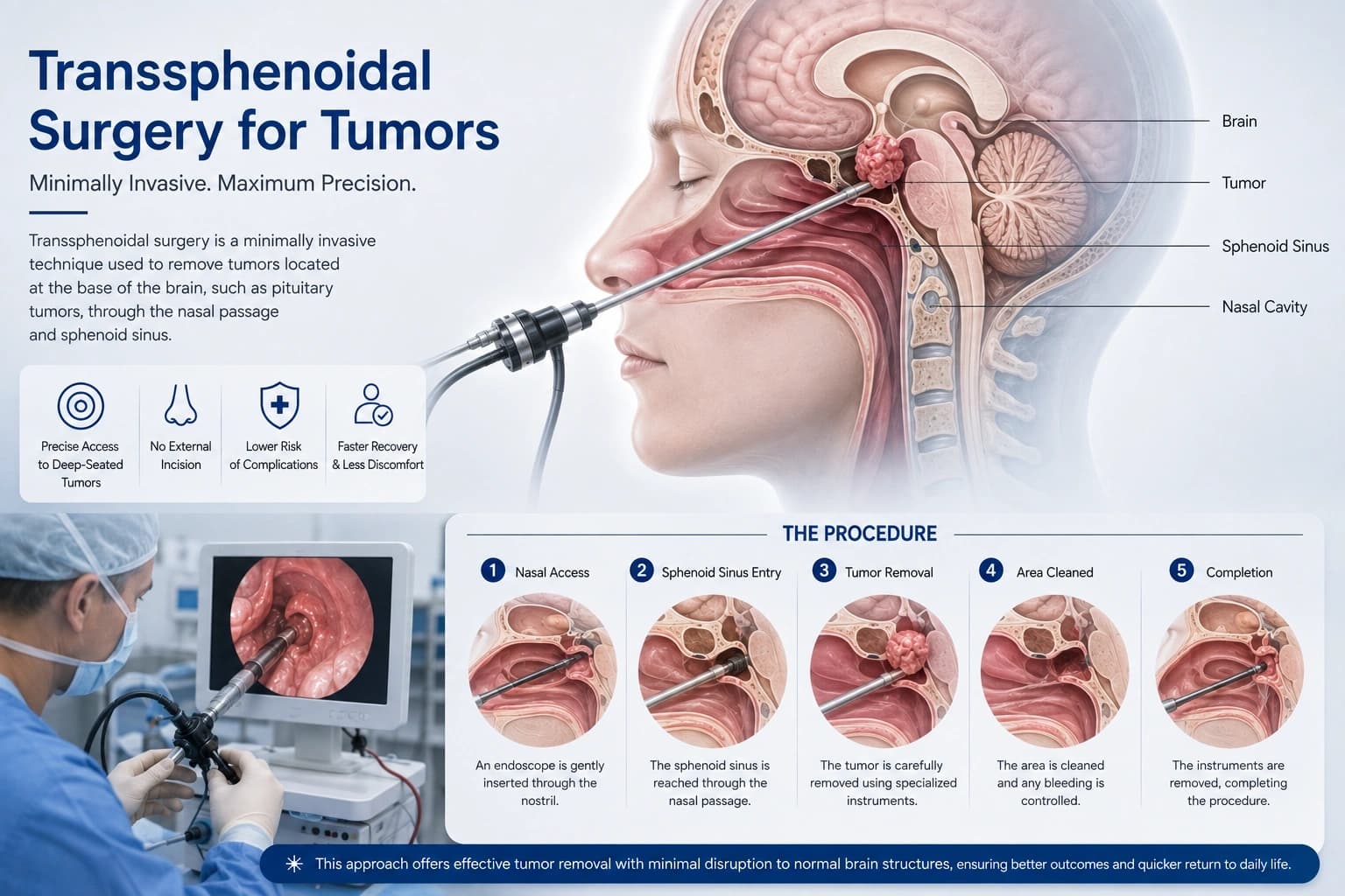
Transsphenoidal Surgery for Tumors
An endoscopic approach through the nasal cavity to remove pituitary adenomas and other tumors at the skull base. This innovative technique avoids facial incisions or brain retraction entirely, offering patients significantly reduced post-operative discomfort, no visible scarring, and a much faster return to daily activities.
Stereotactic Brain Biopsy
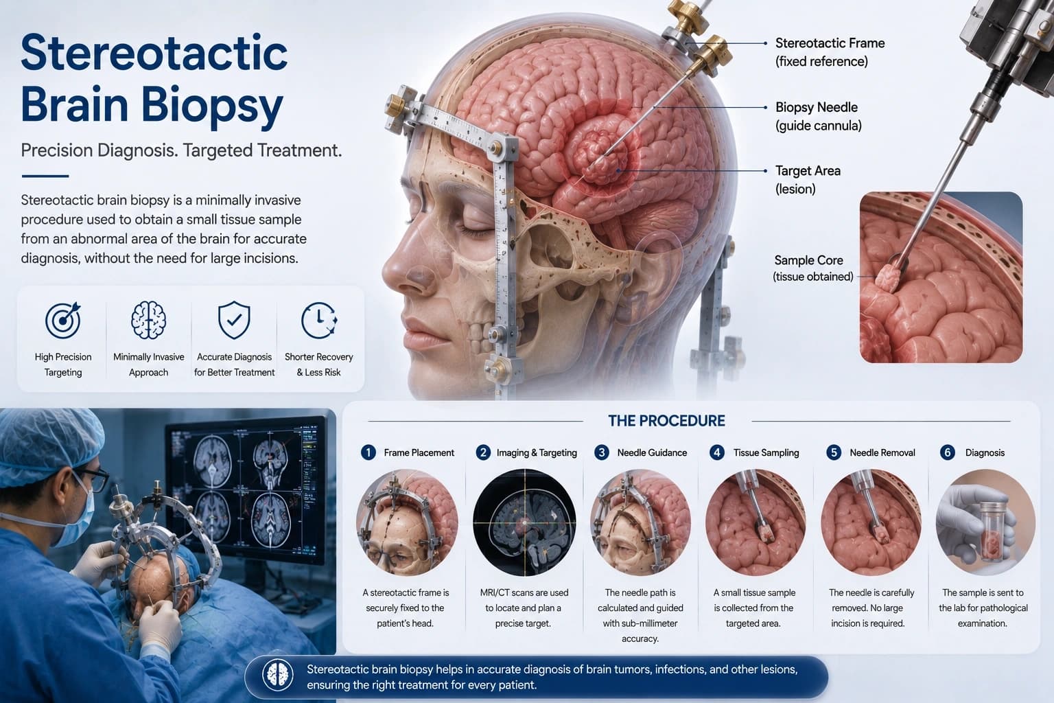
A minimally invasive, image-guided procedure to obtain a small sample of brain tissue for diagnostic purposes. Using highly accurate 3D coordinates based on MRI or CT scans, we guide a specialized needle to deep-seated or eloquent brain lesions to secure a definitive diagnosis with minimal risk to surrounding healthy tissue.
Aneurysm Clipping
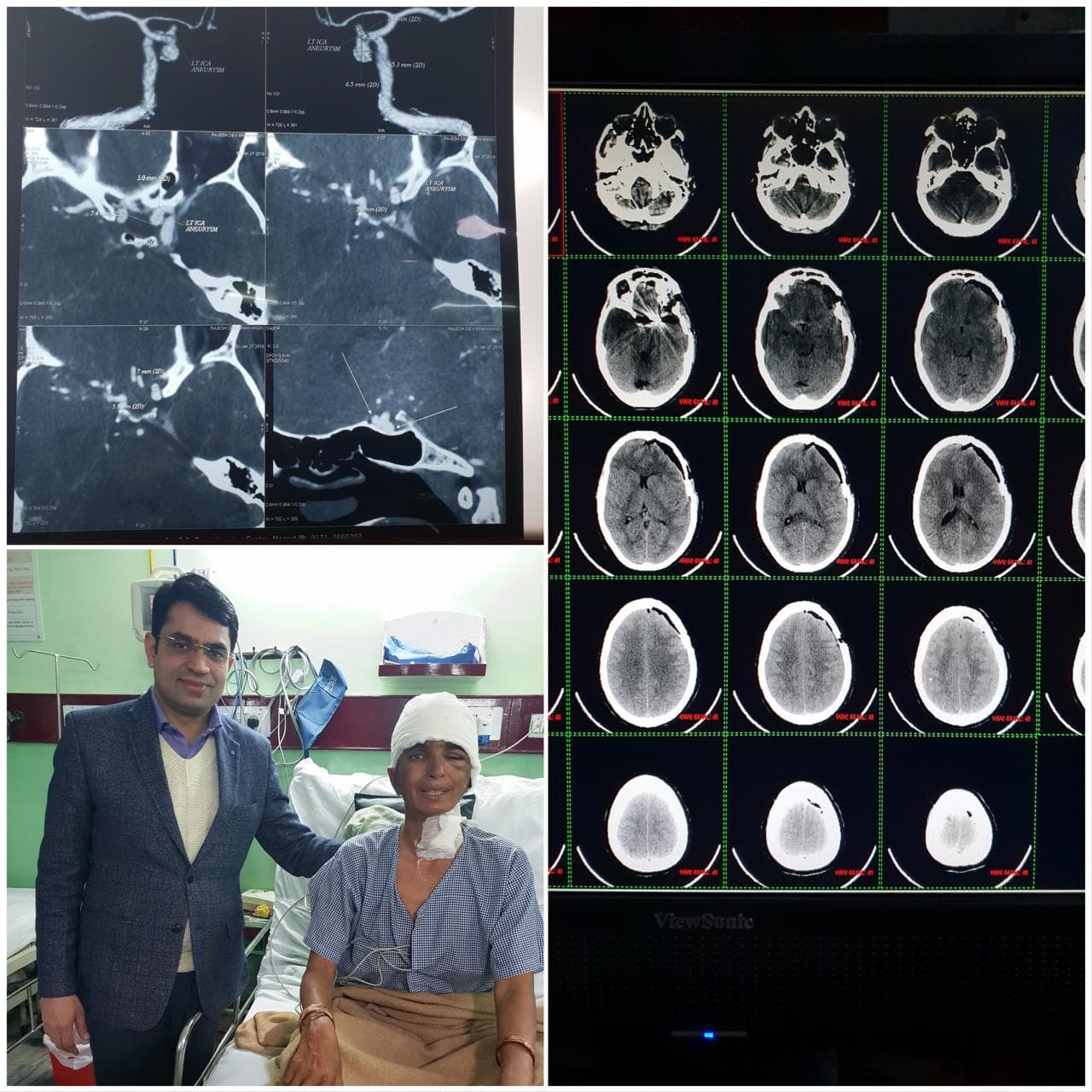
A surgical procedure to treat brain aneurysms. A tiny metal clip is placed at the base of the aneurysm to stop blood flow into it, preventing rupture or re-bleeding. This requires immense precision and advanced microsurgical tools to carefully isolate the aneurysm while preserving the normal parent artery, offering a durable, lifelong solution.
Diagnostic Cerebral Angiography
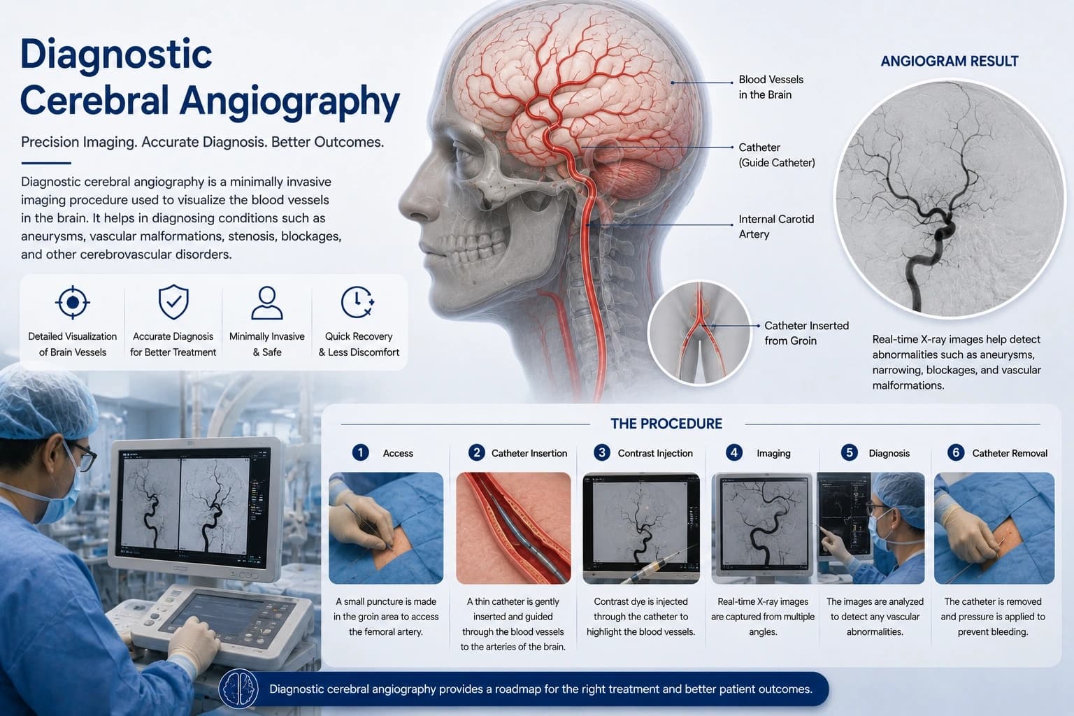
An advanced imaging procedure used to evaluate the blood vessels in the brain. By injecting a contrast dye through a tiny catheter, we obtain real-time, highly detailed maps of the cerebral vasculature. This is considered the gold standard for accurately diagnosing complex aneurysms, AVMs, and identifying critical arterial blockages.
Microvascular Decompression for Trigeminal Neuralgia
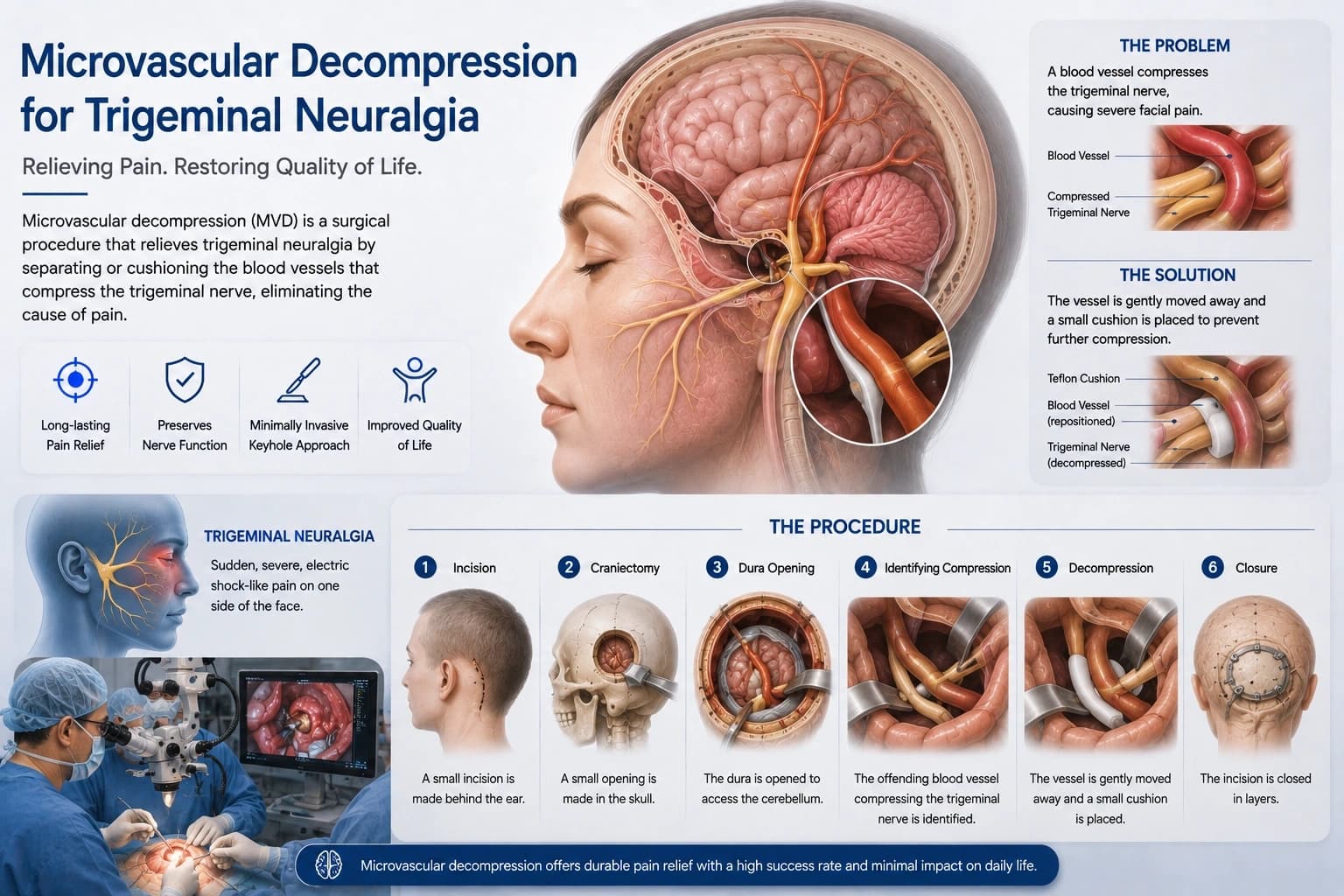
A microsurgical procedure to relieve facial pain by separating the trigeminal nerve from compressing blood vessels. Through a small keyhole opening behind the ear, the offending vessel is carefully moved away from the nerve root and cushioned with a tiny Teflon pad, often providing instantaneous and permanent relief from debilitating facial pain.
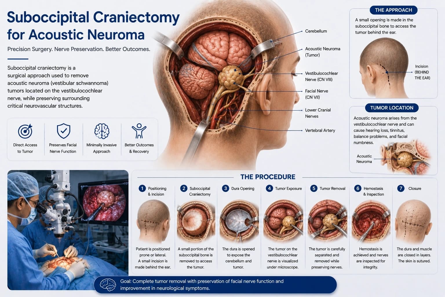
Suboccipital Craniectomy for Acoustic Neuroma
Surgical removal of an acoustic neuroma tumor located near the hearing and balance nerves. Our multidisciplinary skull-base approach utilizes continuous intraoperative cranial nerve monitoring, prioritizing the preservation of facial nerve function and hearing while safely resecting these complex, slow-growing tumors.
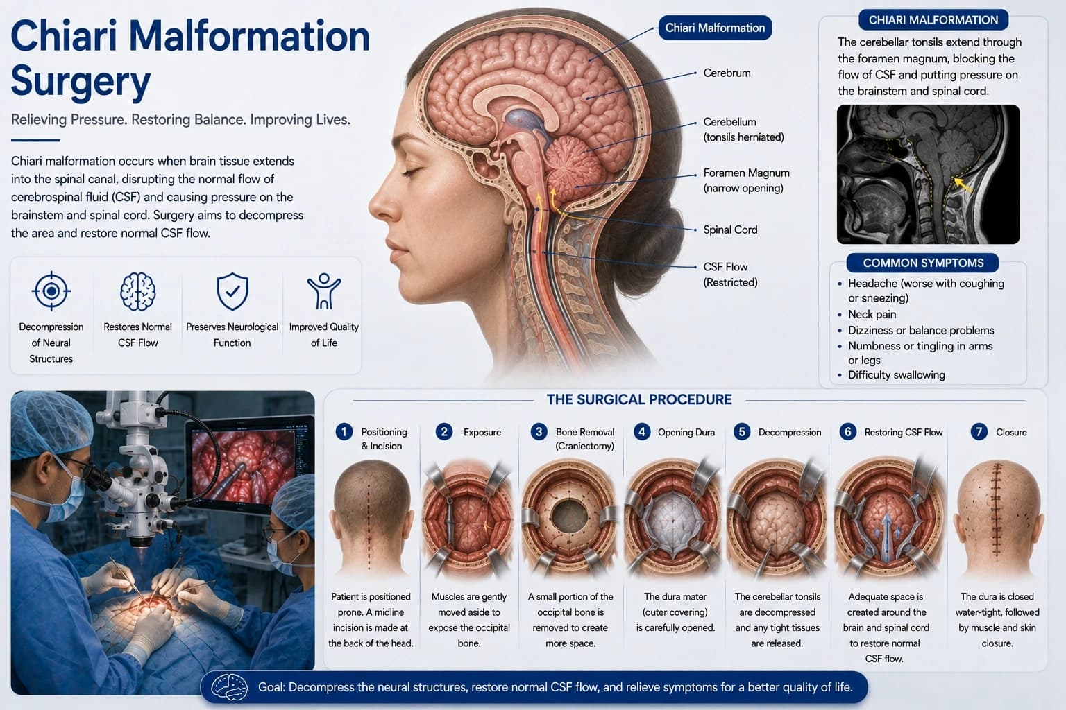
Chiari Malformation Surgery
Surgical decompression to create more space for the cerebellum and relieve pressure on the spinal cord. By removing a small portion of bone at the base of the skull and expanding the protective dural covering, normal cerebrospinal fluid flow is restored, effectively alleviating severe headaches, neck pain, and progressive neurological symptoms.
-Shunt-for-Hydrocephalus.webp&w=3840&q=75)
Ventriculoperitoneal (VP) Shunt for Hydrocephalus
Placement of a shunt system to drain excess cerebrospinal fluid from the brain's ventricles to the abdomen. We utilize advanced programmable valves that can be non-invasively adjusted from the outside, ensuring the precise pressure regulation needed to reverse cognitive decline, gait disturbances, and incontinence.
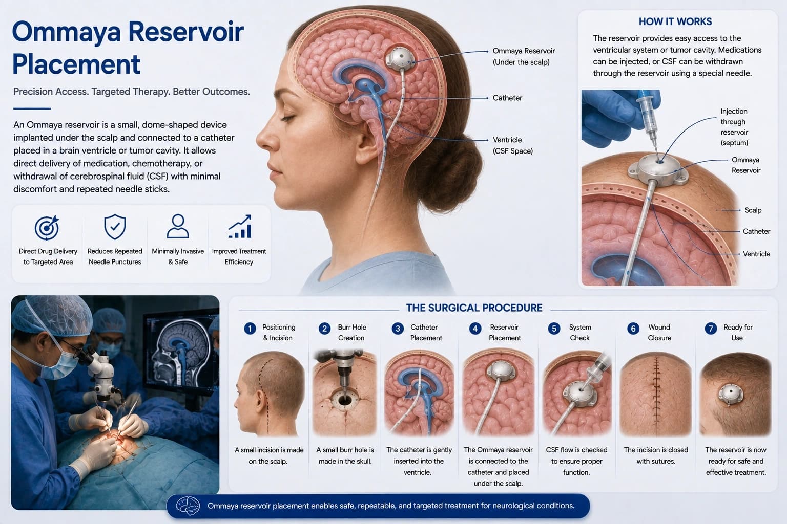
Ommaya Reservoir Placement
Implantation of a specialized device to deliver medication directly into the cerebrospinal fluid or to regularly withdraw fluid. Placed completely beneath the scalp, this reservoir provides a safe, painless, and reliable port for administering long-term chemotherapy or treating complex central nervous system infections.
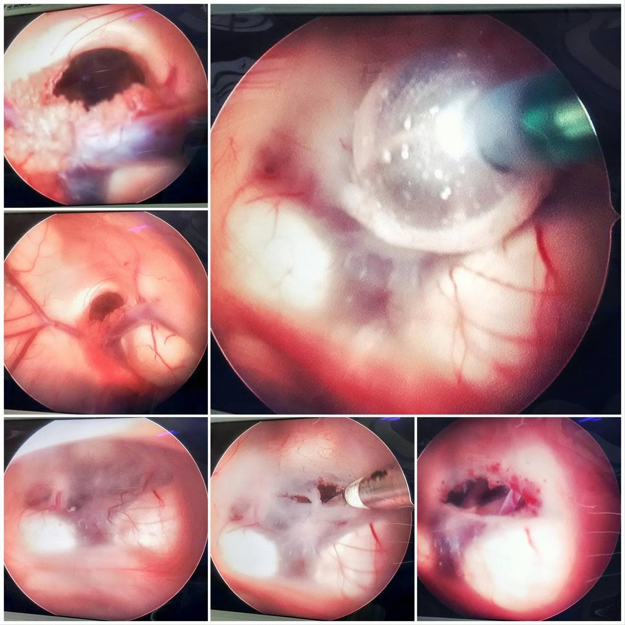
Endoscopic Brain Surgery
A minimally invasive approach used for selected brain tumors, including pituitary adenomas, and conditions such as hydrocephalus. Depending on the condition, an endoscope may be passed through the nose or a small opening in the skull, providing panoramic, high-definition illumination of deep brain structures without the need for extensive traditional craniotomies.
Consult for Cranial Conditions
Expert diagnosis and advanced surgical intervention at Phoenix Spine and Neurosurgery Centre, Meerut.
Book an Appointment